

Angiografía
- Unidad de AngiografíaUnidad de Angiografía
La Unidad de Angiografía de la Clínica Imbanaco Grupo Quirónsalud se encuentra ubicada en el piso 1 de la Sede Principal, cuenta con un ambiente natural, diseñado especialmente para generar más comodidad como a los pacientes.
Está integrada por un grupo de especialistas en terapia endovascular, que desarrollan los más avanzados procedimientos en radiología intervencionista (implante de stent en el sistema arterial), neurorradiología (implantes de stents en el cerebro) y cardiología ( marcapasos y cardiodesfibriladores).
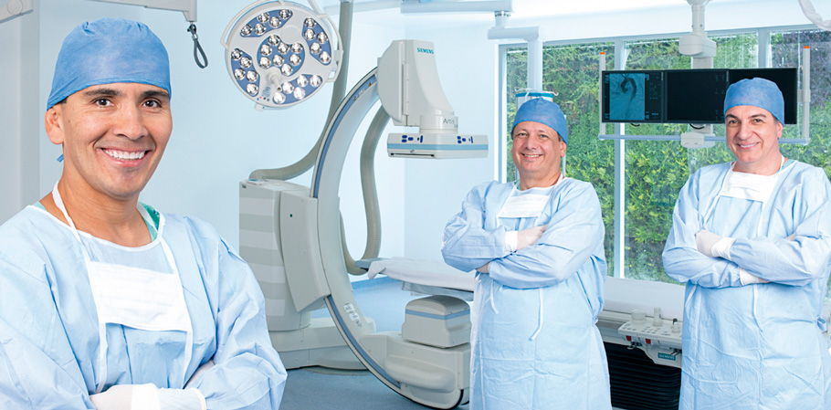
La Unidad de Angiografía de la Clínica Imbanaco Grupo Quirónsalud cuenta con 11 camas de recuperación, además de tener el angiógrafo biplano, que permite ver imágenes del corazón en dos planos, lo que facilita la seguridad y la calidad de la intervención.
- Ventajas y BeneficiosVentajas y Beneficios
- Cuenta con un grupo de especialistas con todo el respaldo académico y científico, además de gran experiencia en la realización de procedimientos angiográficos.
- Cuenta con un personal técnico – asistencial, capacitado y con una gran calidad humana.
- Cuenta con tecnología de vanguardia e instalaciones ampliar adecuadas para prestar una atención segura y confortable.
- Emplea protocolos y estándares internacionales.
- Permanente actualización en técnicas y manejo de insumos, haciendo de la unidad un servicio diferenciados.
- Cuenta con horario 24 horas, para la atención de urgencias.
- ServiciosServicios
Estudios Diagnósticos Invasivos
- Cateterismo cardíaco con determinación de presiones intracavitarias y flujos cardíacos.
- Oximetrías.
- Estudios angiográficos de las cavidades derecha e izquierda del corazón.
- Coronariografía.
- Estudios angiográficos de circulación cerebral.
- Aortogramas torácicos, abdominales y angiografía selectiva de sus ramas.
- Estudios de la circulación periférica: extremidades superiores e inferiores.
- Estudios electrofisiológicos diagnósticos.
- Flebografía.
- Cavografía.
- Angiografía cerebral de 6 vasos.
- Dacriocistografía ocular.
- Angiografía ocular (Ret Cam).
- Punción lumbar.
Procedimientos Terapéuticos Invasivos
- Angioplastia coronaria con implantación de stent.
- Valvuloplastias pulmonar aórtica y mitral con balón.
- Implante de Válvula Aórtica Percutánea.
- Angioplastias de arterias periféricas y cerebrales con implante de dispositivos mecánicos.
- Cierre de ductus con dispositivo endovascular.
- Colocación de balón intraaórtico de contrapulsación.
- Colocación de filtro en vena cava.
- Quimioembolizacion hepática.
- Trombectomía cerebral.
- Trombectomía pulmonar.
- Reparo endovascular.
- Implante de anillo regulador de flujo.
- Implante de valvula pulmonar
- Mitraclip.
- Implante de valvula aortica vía endovascular TAVI.
- Implante de dispositivo WATCHMAN.
Septostomías interauriculares
- Cierre de comunicación interauricular – Foramen oval permeable.
- Implantación de marcapasos unicamerales, bicamerales, monitor de eventos, biventriculares, cardiodesfibriladores y cardiodesfibrilador con resincronizador electrofisiología.
- Cierre de CIV.
- Cierre de ductus.
Electrofisiología
- Estudio electrofisiológico, mapeo y ablación.
- Estudio electroanatómico tridimensional.
- Aislamiento por electroporación.
Terapia Endovascular
- Embolización con coils de aneurismas intra y extracraneales
- Embolización de malformaciones arteriovenosas
- Embolización de tumores
- Quimioterapia intra-arterial
- Trombolisis intra-arterial
- Angioplastia más colocación de stent intra y extracraneal
Procedimientos de Vascular Periférico
- Angioplastia de Miembros Inferiores ó Superiores con Implante de Stent.
- Angioplastia de Arteria Renal más Implante de Stent.
- Reparo Endovascular de Aneurisma de Aorta Torácica y Abdominal.
- Implante de Catéter Translumbar.
- Implante de Catéter para Diálisis.
- Cavernosografía.
Toma de Biopsias guiadas por Angiografía
- Biopsia de hueso.
- Biobsia de corazón.
Manejo de dolor
- Vertebroplastias.
- Neurólisis ganglio de gasser.
- Infiltración y bloqueo de raíces.
- Infiltración y bloqueo de facetas.
- Neurolósis por radiofrecuencia.
- Equipo médicoEquipo médico
Anestesiólogos
- Dr. Juan David Moreno
- Dr. Mario Andrés Varona
- Dra. Alicia Rodríguez
- Dra. Yolima Pupo
- Dr. Luis Eduardo Enriquez
- Dr. Ramiro Moreno
- Dra. Claudia Mercedes Abadía
- Dr. Juan Pablo Vivas
Radiólogos intervencionistas
- Dr. Carlos Alberto González
- Dr. William Escobar Rojas
- Dr. Jorge Andrés Cifuentes
- Dr. Alfredo Pedroza
- Dr. Andrés Ortiz
Manejo del dolor
- Dr. René Alejandro Linares
- Dra. María Mercedes Fajardo
- Dra. Manuela Flórez
- Dra. Paula Rodríguez
Electrofisiólogos
- Dr. Efraín Gil
- Dr. Alberto José Negrete
- Dr. Andrés Gómez
Oftalmólogos oncólogos
- Dra. Carolina Alarcón
- Dra. Ana María Velasco
Hemodinamistas
- Dr. Miled César Gómez
- Dr. Carlos Eduardo Tenorio
- Dr. Julian Andrés Ochoa
Pediátrico
- Dr. Ernesto Vallejo
- Equipo tecnológicoEquipo tecnológico
La Unidad de Angiografía cuenta con novedades tecnológicas disponibles en angiografía como:
- Angiógrafo monoplano marca Siemens: permite obtener imágenes detalladas de los vasos sanguíneos en áreas específicas del cuerpo, lo que ayuda a diagnosticar y planificar tratamientos para diversas condiciones vasculares. Entre los beneficios para el paciente se incluyen la posibilidad de mejorar la precisión de cirugías, obtener diagnósticos precisos y rápidos y, en algunos casos, combinar el diagnóstico con el tratamiento en un solo procedimiento.
- Angiógrafo con el sistema biplano: permite examinar y tratar problemas dentro del corazón, cerebro, grandes arterias y venas con mayor seguridad. Tecnología con una visión 360° y reconstrucción de la imágenes 3D Y 4D.
Ofrece múltiples beneficios para el paciente, principalmente en la precisión diagnóstica y la posibilidad de intervenciones mínimamente invasivas, en menos tiempo y con menos uso de medios de contraste. Permite una visualización detallada y tridimensional de los vasos sanguíneos, facilitando la identificación de problemas como estrechamientos, obstrucciones o aneurismas y posibilitando la planificación precisa de tratamientos como la colocación de stents o la cirugía. - Polígrafo Cardiotek Ep – tracer y Ep – Work Mate: un paciente que se someta a un estudio con este equipo puede obtener beneficios relacionados con la evaluación de su estado de salud cardiovascular y la detección temprana de posibles problemas.
Estos estudios permiten analizar la actividad eléctrica del corazón, identificar arritmias y evaluar la respuesta del sistema cardiovascular a diferentes estímulos;, lo que puede ayudar a los médicos a diagnosticar y tratar enfermedades cardíacas de manera más efectiva. - Ablación por radiofrecuencia (ARF) con el Generador de Radiofrecuencia IBI T7 y IBI T11: para la realización de procedimientos a nivel diagnóstico y terapéutico en la especialidad de electrofisiología. Se usa para el tratamiento del dolor. Puede proporcionar alivio del dolor crónico, una recuperación más rápida y menos invasiva en comparación con la cirugía y la posibilidad de repetir el procedimiento si el dolor regresa.